
Figure 8. Electronmicrographs of bushy cell dendrites in cross section.
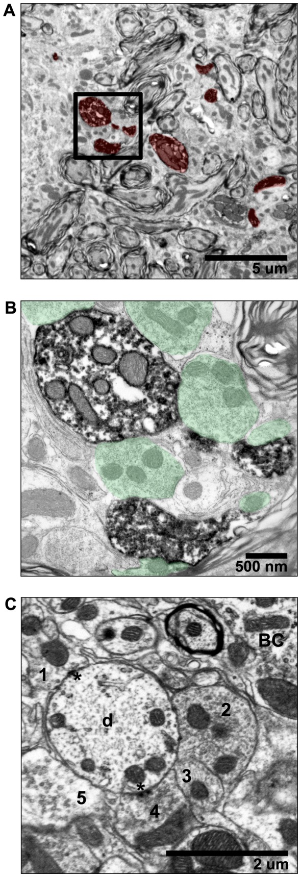
(A) Multiple pieces of a labeled dendrite are visible in the neuropil surrounding bushy cells. (B) Bushy cell dendrites receive numerous bouton-like synaptic terminals (in green). This image shows terminals contacting the labeled dendrites in the boxed area in A. (C) Synaptic terminals (1–5) contacting an unlabeled dendrite (d) are shown so that ultrastructural details are visible. PSDs are marked with asterisks. Bushy cell (BC) body is indicated.