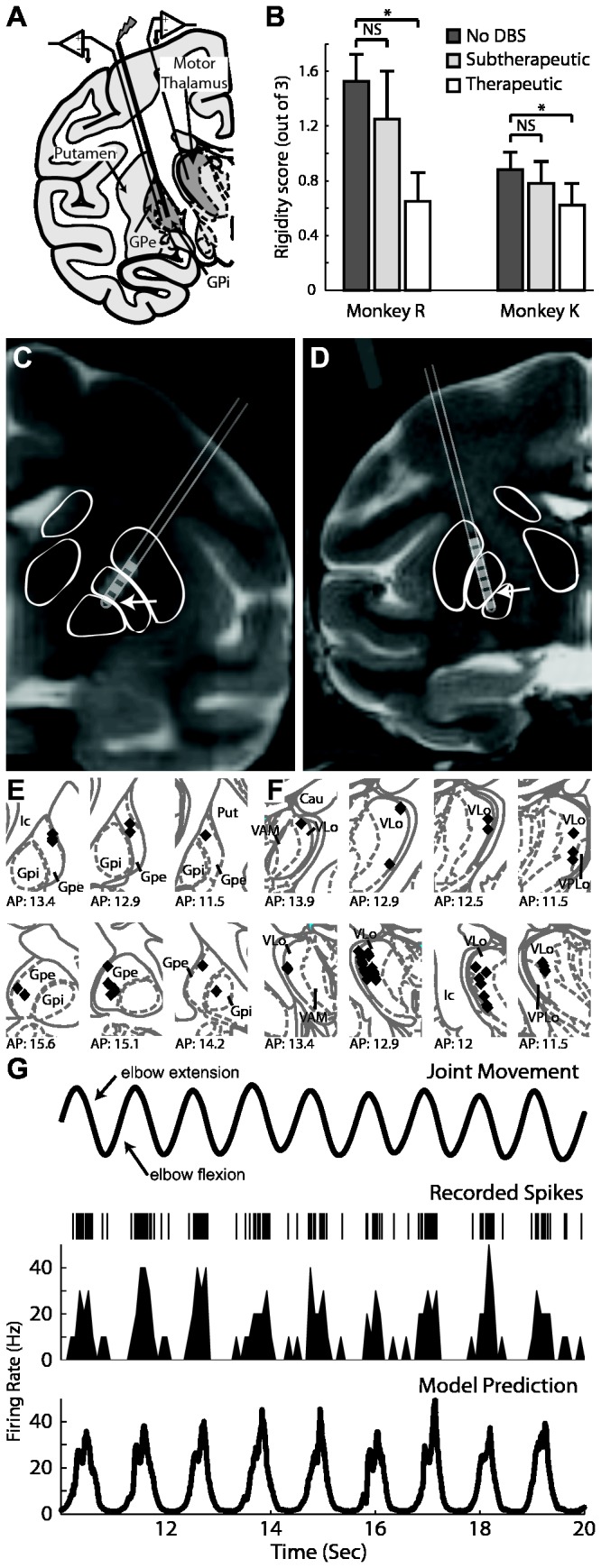
Figure 1. Experimental design used to investigate the effects of GP-DBS on encoding of joint kinematics through the pallidofugal pathway.

A: Microelectrode recordings were performed in regions of the globus pallidus and thalamus with spike activity that was responsive to passive joint movement. B: Results of experimenter-blinded muscle rigidity scoring for both monkeys at three DBS settings. C and D: Co-registration of pre-operative MRI and post-electrode implantation CT showing DBS electrode location for monkey R (C) and K (D). E and F: Localization of recorded cells obtained from stereotactic navigation software and overlaid on corresponding atlas plates for monkey R (top) and K (bottom) for both the pallidum (E) and the thalamus (F). G: A generalized linear model (GLM) accounting for position, velocity, and acceleration of the joint movement was applied to determine the correlation between kinematics of the joint movement (top row) and spike activity (2nd row: spike raster, 3rd row: corresponding rate histogram). Bottom row shows the GLM prediction of firing rate.
