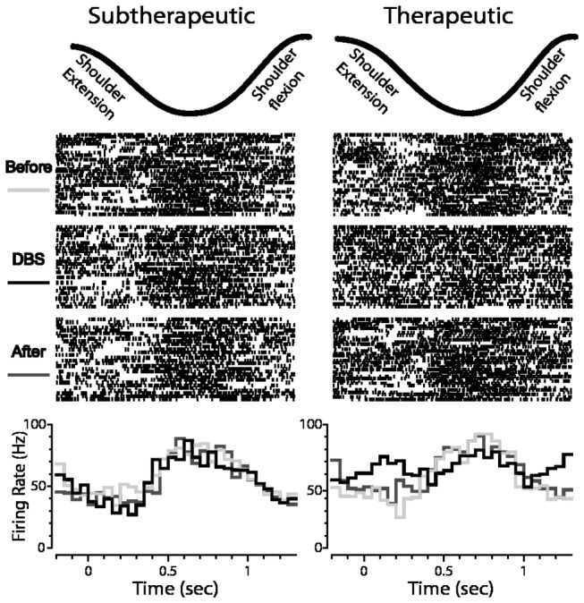
Figure 5. Neuronal encoding of joint movement during subtherapeutic and therapeutic DBS in globus pallidus.

Shown is an example of the response of a cell to shoulder flexion/extension before, during and after subtherapeutic DBS (left) and therapeutic DBS (right) (top: motion capture data of the joint movement; middle: corresponding raster plots triggered to the beginning of each movement cycle; bottom: PETHs showing responses before, during, and after DBS).
