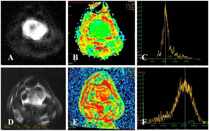
Figure 2. Osteosarcoma of the distal femur in a 24-year-old man with good response.
(A∼C) DWI map, ADC map and ADC histogram before neoadjuvant chemotherapy. The signal intensity of tumor on DWI map was high. The ADC value of the whole tumor was 1.12×10⁻³ mm2/s with a green area. The ADC histogram was high and sharp. (D∼F) DWI map, ADC map and ADC histogram after neoadjuvant chemotherapy. The signal intensity of tumor on DWI map was decreased. The ADC value of the whole tumor was increased to 1.99×10⁻³ mm2/s with a subtotal red area. The ADC histogram was short, wide and moving to the right of the coordinate. Tumor necrosis rate of 92% was confirmed by postoperative pathological evaluation. The effectiveness of neoadjuvant chemotherapy was good.

