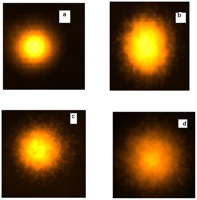
Figure 10. Comet assay.

Images of treated lymphocyte nuclei (a) negative control; (b) in the presence of 3 µl of methyl methane sulfonate (25 µg/ml) as positive control (c) Hb aggregates formed at 70% glyoxal (d) glycated Hb at 30% glyoxal on day 20. The protein concentration was 50 µg.
