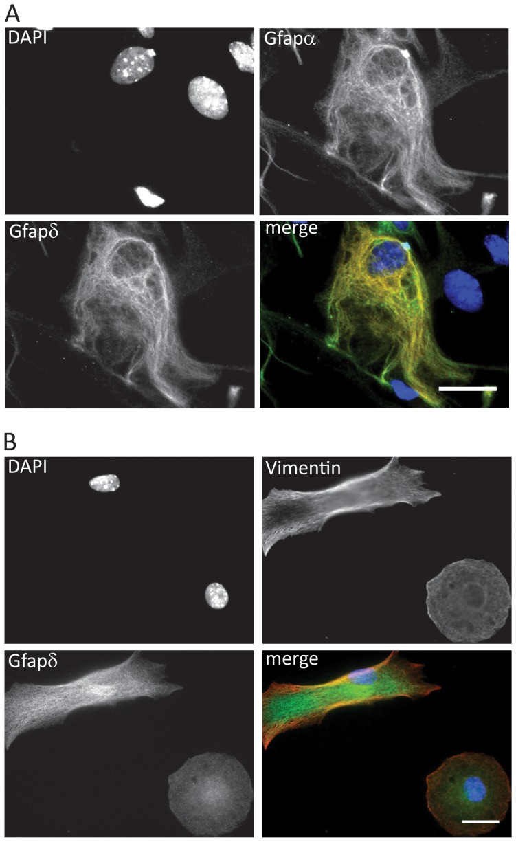
Figure 4. Immunofluorescence analysis of Gfapδ.
(A) Co-localization analysis of Gfapα and Gfapδ. Mouse primary astrocytes were stained with primary Gfapδ antibody and Gfapα antibody. The nuclei were counterstained with DAPI. Merged image is included with Gfapα labeled green, Gfapδ labeled red and DAPI labeled blue. (B) Gfapδ and Vimentin have partial co-localization. Mouse primary astrocytes were stained with primary Gfapδ antibody and Vimentin antibody. The nuclei were counterstained with DAPI. Merged image is included with Vimentin labeled red, Gfapδ labeled green and DAPI labeled blue. Scale bar 10 µm.

