
Figure 2. Enhanced stem cell recruitment around chemokine loaded implants.
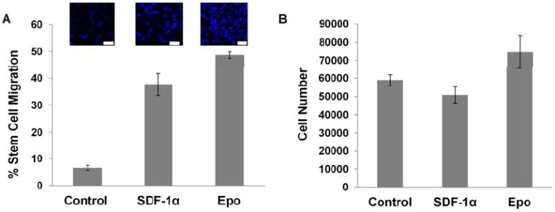
(A) Recruitment after 24 hours of injected NIR dye labeled MSCs to chemokine SDF-1α and Epo loaded microbubble scaffolds was imaged and quantified. MSC recruitment was compared based on (B) CD105+/ CD45-CD34-CD56-, (C) CD146+/ CD45-CD56- and (D) Stro-1+/ CD34-CD45- MSC markers using immunofluorescence labeling and quantified. (n=4; Mag 400X; significance of mean values with respect to control tested at p<0.05, *).