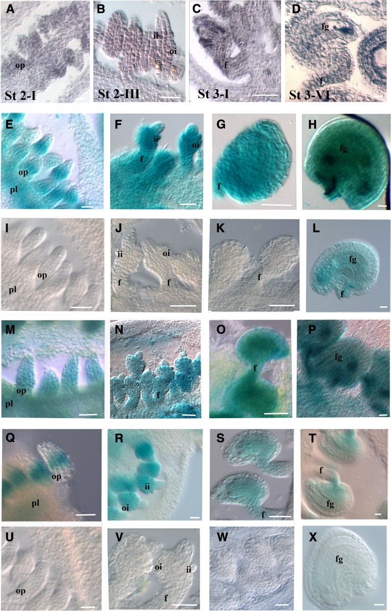
Figure 2.
VDD Expression and Promoter Analysis during Ovule Development.
(A) to (D) In situ hybridization analysis of VDD during ovule development. Ovule development stage 2-I (A), stage 2-III (B), 3-I (C), and stage 3-VI (D) (these stages should be used as reference for the next lines).
(E) to (H) proVDD:GUS transgenic plants showed a similar expression pattern as observed by the in situ hybridization experiment.
(I) to (L) GUS expression in ovules of proVDDm1:GUS lines.
(M) to (P) GUS expression in ovules of proVDDm2:GUS lines.
(Q) to (T) GUS expression in ovules of proVDDm2:GUS lines.
(U) to (X) Absence of GUS expression as observed in the proVDDm1-2:GUS. Absence of GUS expression was also observed in proVDDm1-3:GUS, proVDDm2-3:GUS, and proVDDm1-2-3:GUS lines.
pl, placenta; op, ovule primordium; f, funiculus; ii, inner integument; oi, outer integument; fg, female gametophyte. Bars = 20 µm.

