Figure 1.
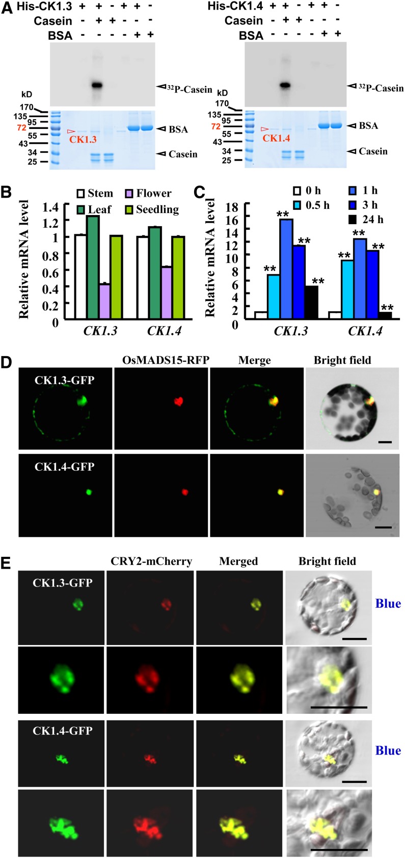
CK1.3 and CK1.4 Are Expressed in Various Tissues and Encode Active Casein Kinases.
(A) In vitro kinase assays by [γ-32P]ATP autoradiography indicated that recombinant CK1.3 and CK1.4 display CK1 activity and specifically phosphorylate casein in vitro (top panel; BSA was used as control). Coomassie blue (CBB) staining showed that equal quantities of protein (10 μg; bottom panel) were loaded.
(B) qRT-PCR analysis revealed the expression of CK1.3 and CK1.4 in stems, leaves, flowers, and seedlings. Expression level of CK1.3 or CK1.4 in stems was set as 1.0. Error bars represent se. The experiments were repeated three times.
(C) qRT-PCR analysis revealed the induced expression of CK1.3 and CK1.4 under blue light. Seedlings were grown under a 3 µmol/m2/s fluence rate of blue light for 0.5, 1, 3, or 24 h. Expression level of CK1.3 or CK1.4 in the dark was set as 1.0. The experiments were repeated three times (**P < 0.01, n = 3).
(D) Transient expression analysis using Arabidopsis protoplasts revealed that both CK1.3-GFP and CK1.4-GFP fusion proteins are mainly localized in the nucleus. A nucleus-localized OsMADS15-RFP fusion protein (Zhang and Xue, 2013) was used as a positive control. Protoplasts were observed after incubation for 9 h. Bars = 20 μm.
(E) CK1.3-GFP and CK1.4-GFP fusion proteins are accumulated in nuclear bodies under blue light treatment. Colocalization analysis by transiently expressing CK1.3-GFP and CK1.4-GFP in Arabidopsis protoplasts expressing CRY2-mCherry indicated the colocalization of CK1.3-GFP and CK1.4-GFP with CRY2-mCherry (enlarged at bottom panels). Protoplasts were exposed to blue light (10 µmol/m2/s) for 5 min before observation. Bars = 20 μm.

