Figure 5.
CK1.3 and CK1.4 Interact with and Phosphorylate CRY2, Which Is Stimulated by Blue Light.
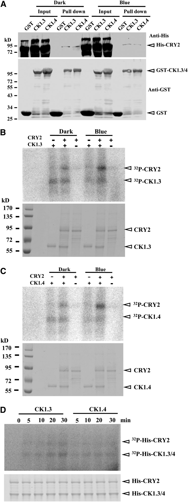
(A) In vitro GST pull-down assay revealed the interaction of CK1.3 and CK1.4 proteins with CRY2 under dark or blue light exposure (10 µmol/m2/s). The quantitative assay of the input proteins is shown. GST was used as a negative control (bottom panel). GST-CK1.3 and GST-CK1.4 were expressed and purified from E. coli and His-CRY2 from insect cells. CK1.3, CK1.4, and CRY2 were detected by a His tag antibody (top panel) or a GST tag antibody (bottom panel), respectively. Positions of CK1.3, CK1.4, and CRY2 are highlighted by arrowheads.
(B) and (C) In vitro kinase assay indicated that phosphorylation of CRY2 by CK1.3 (B) and CK1.4 (C) is stimulated by blue light exposure. All the reactions were initiated by adding kinases under red light and then transferred to dark or blue light (10 µmol/m2/s) for 3 h. Kinase activity was detected by [γ-32P]ATP autoradiography (top panels), and equal input of His-CK1.3, His-CK1.4, and His-CRY2 (10 μg) was shown by Coomassie blue staining (bottom panels).
(D) In vitro kinase assay by [γ-32P]ATP autoradiography revealed that CK1.3 and CK1.4 rapidly phosphorylated CRY2 under blue light (top panel). Purified recombinant His-CRY2 protein from insect cells was used for the assay, and all reactions were initiated by adding CK1.3 or CK1.4 under red light before blue light exposure. Blue light (10 µmol/m2/s) treatment was applied for 0, 5, 10, 15, or 30 min. The input of His-CRY2, His-CK1.3, and His-CK1.4 were detected by Coomassie blue staining (bottom panel).

