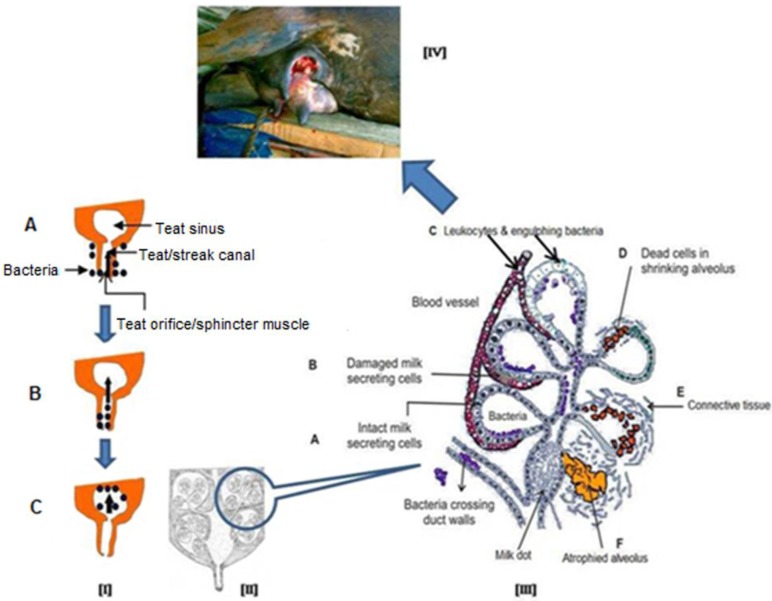
Figure 3.
Schematic representation of process of intramammary infection and subsequently damage to mammary gland. [I] Bacteria present on the surface of mammary gland (udder, A) and enter into the teat canal (B) after getting opportunity and finally setup the infection in mammary gland (C), [II] A longitudinal diagram of normal mammary gland, [III] After getting bacterial infection, cellular defence mechanism become active and phagocytic cells (from blood) effort to engulf and kill the bacteria, phagocytosis by-products and release of bacterial toxins damage to the secretory mammary epithelial cells and finally cause fibrosis of mammary gland (A to F), [IV] Final outcome of acute and/or chronic mastitis.

