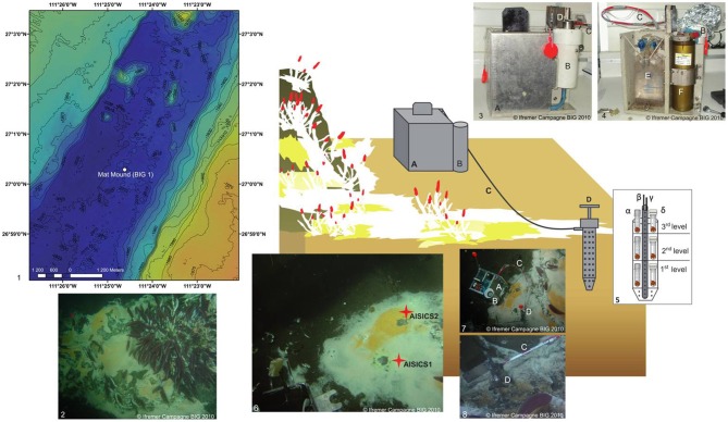
Figure 1.
Schematic diagram illustrating the deployment of the in situ AISICS module at Mat Mound site. (1) Bathymetric map showing the location of Mat Mound site in the Southern Trough of the Guaymas Basin; (2) Mat Mound site exhibiting microbial mats and macro-fauna dominated by Riftia tub tube worms (Siboglinidae); (photo taken with the submersible Nautile during the BIG cruise, Dive 1745); (3) AISICS module covered by its lid before its deployment; (4) AISICS module without its lid before its deployment; (5) Diagram illustrating the internal structure of the incubator with biotic (α) and abiotic (β) mini-colonizers distributed per floor; the central titanium sheath containing the Micrel temperature sensor (γ) and the fluid sample probe (δ) hosted in a titanium sheath are placed in the middle of the incubator and are connected to the instrumented base; (6) The deployment site of AISICS1 and 2; (7) The deployed AISICS1 (photo was taken with the submersible Nautile during the BIG cruise, Dives 1745); (8) The deployed AISICS2 (photo was taken with the submersible Nautile during the BIG cruise, Dives 1763); (A) instrumented module; (B) cylindrical insulated chamber; (C) sampling pipes and temperature probe; (D) incubator; (E) sampling pouches, and (F) electronics.

