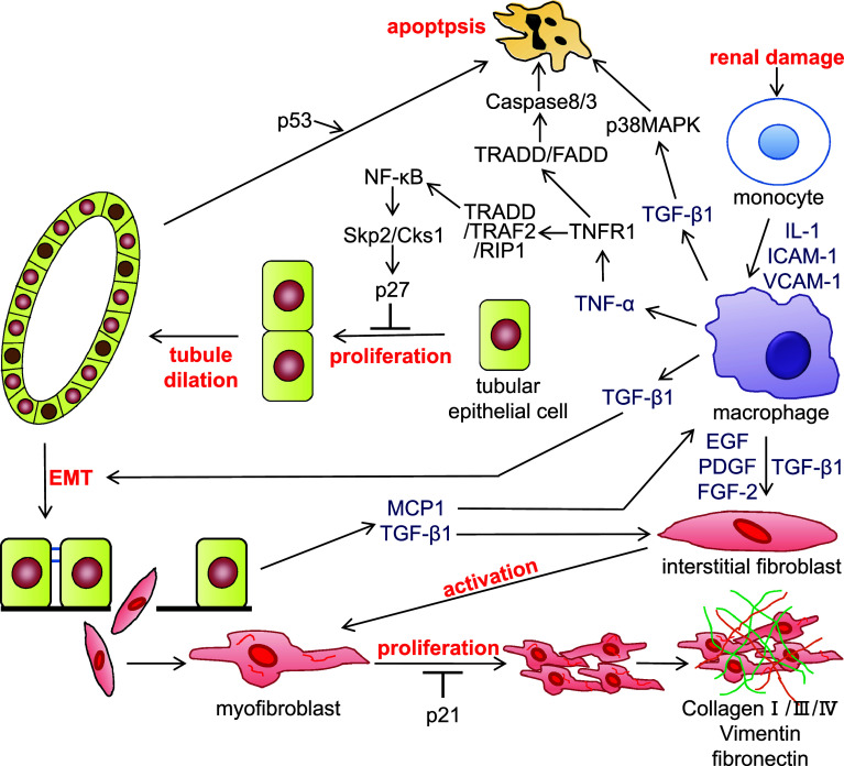
Fig. 1.
The signal transduction pathways involved in the progression of chronic nephropathy. Following renal damage, infiltrated macrophages in the tubulointerstitium release cytokines such as tumor necrosis factor-α (TNF-α) and transforming growth factor-β1 (TGF-β1). TNF-α binds to TNF receptor 1 (TNFR1) and forms a complex with TNFR-associated death domain (TRADD), TNF associated factor 2 (TRAF2), and receptor interaction protein 1 (RIP1). This complex activates nuclear factor (NF)-κB, which induces Skp2 and Cks1. Upregulation of Skp2/Cks1 promotes p27 degradation in tubular epithelial cells, allowing proliferation of tubular epithelial cells and tubule dilation following the increase of tubular epithelial cell number. The tubular epithelial cells undergo epithelial-mesenchymal transition (EMT) by stimulation of TGF-β1, and the resulting fibroblasts migrate to the tubulointerstitium. Cytokines including TGF-β1 activate fibroblasts; activated myofibroblasts produce extracellular matrix components, such as collagen, vimentin, and fibronectin. Meanwhile, TNF-α and TGF-β1 induce tubular epithelial cell apoptosis. IL-1 interleukin-1, ICAM-1 intercellular adhesion molecule-1, VCAM-1 vascular cell adhesion molecule-1, EGF epidermal growth factor, PDGF platelet-derived growth factor, FGF-2 fibroblast growth factor-2, FADD Fas-associated death domain protein, MAPK mitogen-activated protein kinase

