Fig. 5.
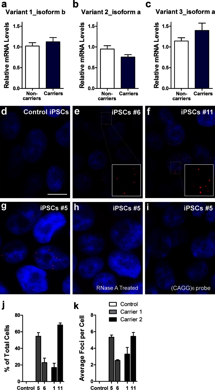
C9ORF72 repeat expansions form RNA foci in iPSCs. Expression levels of C9ORF72 variant 1 (NM_145005.5, isoform b) (a), variant 2 (NM_018325.3, isoform a) (b) and variant 3 (NM_001256054.1, isoform a) (c) in iPSC lines from two non-carriers and two expanded repeat carriers were assessed by qRT-PCR. Fluorescence in situ hybridization (FISH) analysis was done on control iPSC line 20 (d), carrier 1 line 6 iPSCs (e), carrier 2 line 11 iPSCs (f) using a cy3-conjugated (GGCCCC)4 probe. RNA foci (red) were found in the nucleus (blue) of carriers 1 and 2 but not in control cells. Treatment of iPSCs with RNase A after fixation leads to loss of foci (h), indicating that the foci are indeed made of RNA. Representative images of iPSCs from carrier 1 (line 5) that were left untreated (g) or treated with RNase A (h) for 20 min at room temperature. Red RNA foci containing GGGGCC repeats; blue nuclei (DAPI). Cells did not show foci when a Cy3-conjugated (CAGG)6 probe was used as the negative control probe (i). Scale bar 10 μm. Quantifications of the percentage of iPSCs displaying foci (j) and the average number of foci per cell (k) are shown as mean ± SEM

