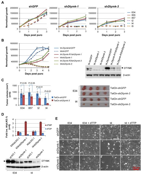
Figure 2. Dtymk is the synthetic lethal target of Lkb1 loss.
A, Lkb1-wt (634, 855, and 857) and Lkb1-null (t2, t4, and t5) cells were transduced with the indicated shRNA for 2 days and then plated into 96-well plates at 2000 cells/well in 150 μl medium with 3 μg/ml puromycin (puro). Viable cells were measured daily using Promega’s CellTiter-Glo Assay. The data represent mean ± SD for 3 replicates.
B, Lkb1-null t4 cells were first transduced with pLenti6-Dtymk, pLenti6-Dtymk-R1, or pLenti6-Dtymk-R3, and selected with blasticidin. The blasticidin-resistant cells were pooled and further transduced with the indicated shRNA for 2 days and then plated for proliferation assay as described in A. The data represent mean ± SD for 3 replicates.
C, 1×10⁶ Lkb1-wt (634 and 857) and Lkb1-null (t2 and t4) cells transduced with the indicated shRNA were implanted into athymic nude mice for 3 weeks. Tumor volume (mm3) was calculated as (length × width2)/2. The data represent mean ± SD for 4 mice. Lkb1-wt 634 and Lkb1-null t4 tumors with the indicated shRNAs were shown.
D, Graph of dTMP and dTDP levels in Lkb1-wt 634 and Lkb1-null t4 cells transduced with the indicated shRNA for 3 days. The data represent mean ± SD for 6 replicates. Expression of DTYMK in these cells at the time of metabolite extraction was determined by Western blotting.
E, Morphology of Lkb1-wt 634 and Lkb1-null t4 cells transduced with the indicated shRNA and then cultured in medium with or without additional 100 μM dTTP for 4 days.

