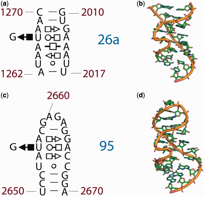
Figure 3.
Loop E motifs: Helix 26a and Helix 95 of the 23S rRNA of E. coli. (a) The 2° structure and (b) the 3D structure of the rRNA that was traditionally represented as single-stranded, adapted from Leontis et al. (25,28). The symbols in the fragments of the 23S rRNA 2° structure represent non-Watson–Crick base pairs: circles correspond to the Watson–Crick edges, squares to the Hoogsteen edges, triangles to the sugar edges, the open symbols indicate trans basepairs and closed symbols, cis basepairs. (c) The 2° structure and (d) the 3D structure of the sarcin-ricin loop (Helix 95). A comparison the top and bottom panels illustrates the extent of 2° and 3D conservation of the loop E motif.

