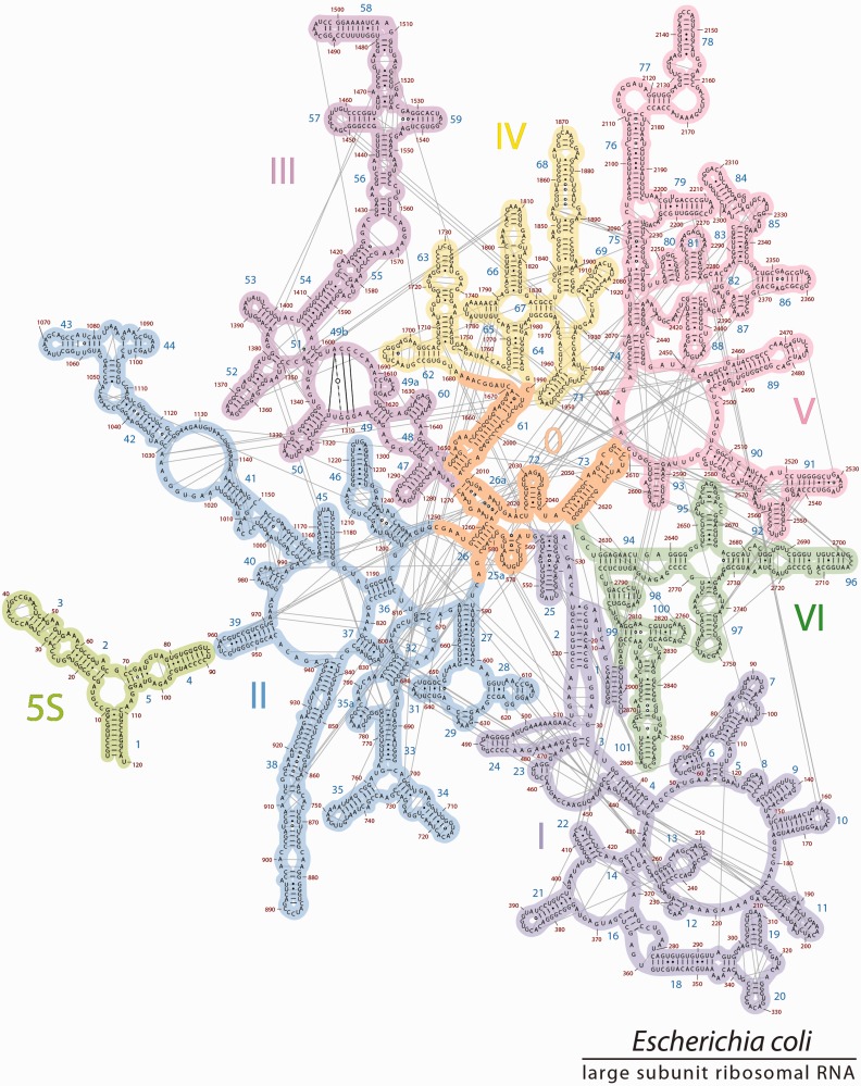
Figure 6.
The mapping of all base pairing interactions onto 2° structure3D. Nucleotides that are base paired in the 3D structure of the ribosome are connected by lines in the 2° structure here. The 5S rRNA is placed in proximity to Helix 39 to reflect their relative locations in 3D space. The interactions between 23S and 5S rRNAa are illustrated in Supplementary Figure S4. Domain 0 is stabilized by many base-pairing interactions. The coloring scheme of the domains is the same as in Figure 5. The most frequent subtypes of base pair interactions [cWW, tWW, tSS and cSS, defined by Leontis (26)] are illustrated in Supplementary Figure S6.

