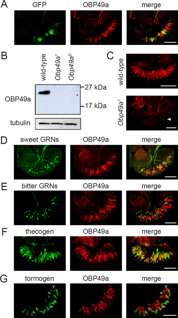
Figure 3. Expression of OBP49a in the labellum.
(A) Co-expression of the Obp49a reporter and anti-OBP49a. Obp49a1/UAS-mCD8::GFP labella were stained with GFP and OBP49a antibodies. (B) Western blot using OBP49a antibodies. Extracts were prepared from wild-type, Obp49a1, and Obp49aD labella, and the blot was probed with anti-OBP49a. The blot was also probed with anti-tubulin as a loading control. (C) Staining of labella from wild-type (top) and Obp49a1 flies with anti-OBP49a (bottom). The arrowhead indicates the position of the thecogen cells in the Obp49a1 mutant labellum. (D–G) OBP49a was expressed in thecogen cells. The cellular distribution of OBP49a was examined by comparing the spatial distribution of cell-type-specific markers with anti-OBP49a. (D) Gr5a-GAL4/UAS-mCD8::GFP (sweet GRNs). (E) Gr66a−/−GFP (bitter GRNs). (F) nompA-GAL4/UAS-mCD8::GFP (thecogen). (G) ASE5-GFP (tormogen). Scale bars represent 50 µM

