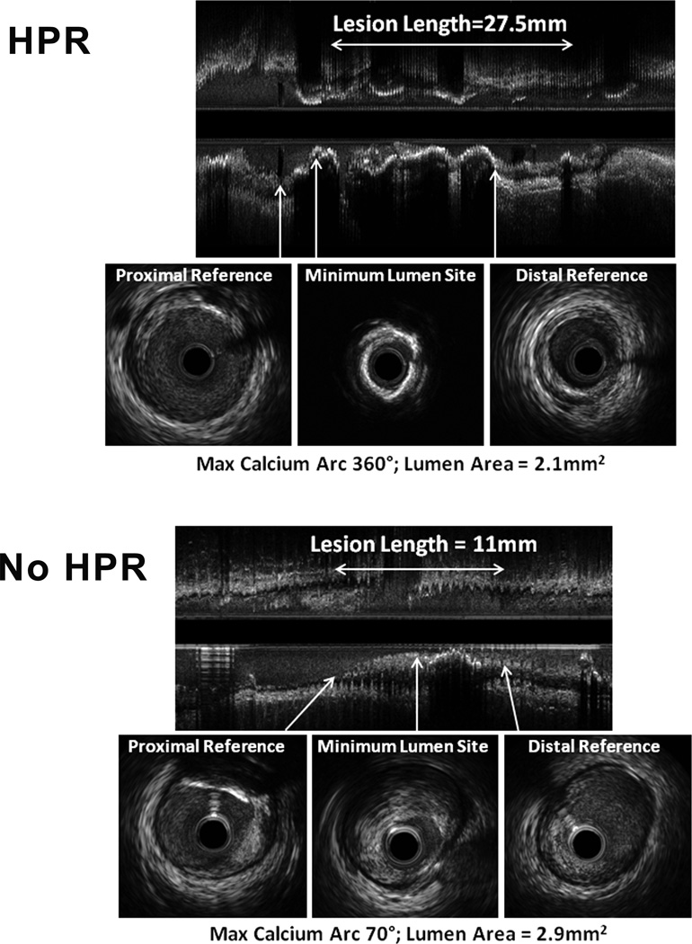
Figure 1. Illustrative volumetric IVUS images from ‘HPR’ and ‘No HPR’ patients.
Upper panel shows an example of a ‘HPR’ patient. This long lesion (27.5 mm from proximal to distal reference site) contains diffuse calcification (note the acoustic shadows beyond regions of calcification) with a maximum calcium arc of 360° at the minimum lumen area (MLA) site. Lower panel shows a ‘No HPR’ patient. While not intended to be representative, the lesion length was shorter (11 mm from proximal to distal reference site) with non-calcified plaque at the MLA site; the maximum arc of calcium (70°) was located at the proximal reference. Both lesions had lumen areas at the lesion site of < 3.0 mm2, considered to be highly hemodynamically significant.

