
Figure 1.
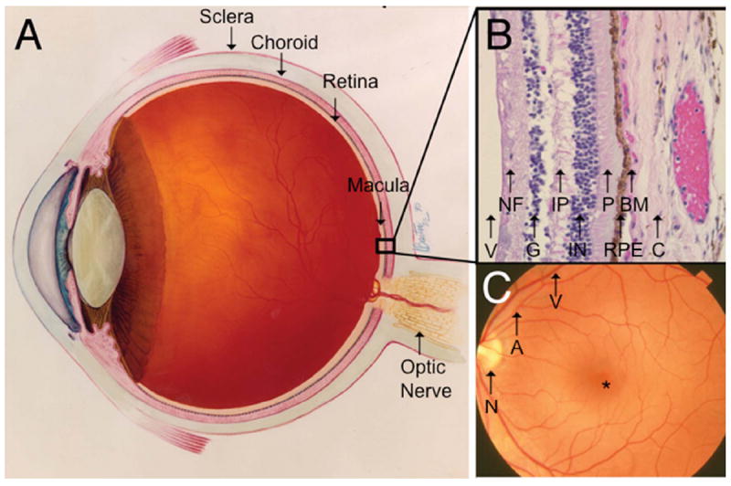
Ocular anatomy relevant to AMD. A. Cross-section of an eye showing the retina and neighboring anatomic structures. B. Histological cross-section of the retina moving inward along the path of incident light include the vitreous (V); Nerve fiber layer (NF) consisting of axons of the third neuron; Ganglion cell layer (G) consisting of the cell nuclei of the multipolar ganglion cells of the third neuron; Inner plexiform layer (IP) consisting of synapses between the axons of the second neuron and dendrites of the third neuron; Inner nuclear layer (IN) containing cell nuclei of the bipolar nerve cells on the second neuron, horizontal cells, and amacrine cells; Photoreceptors (P) consisting of rods and cones; Retinal pigment epithelium (RPE) consisting of a single cubic layer of heavily pigmented epithelial cells; Bruch’s membrane (BM) the matrix separating the RPE from the choroid; Choroid (C), the vasculature between the retina and sclera. C. Fundoscopic image showing the macula located between vascular arcades including the retinal artery (A) and vein (V) with the fovea (*) at the center, approximately 3 mm temporal to the optic nerve (N). Images courtesy of the National Eye Institute.