Figure 1.

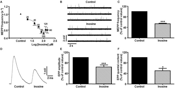
Inhibitory effect of inosine on spontaneous and evoked ACh release at the mouse NMJ. (A) Effect of inosine on MEPP frequency (s−1) as a function of its concentration. Each point represents mean ± SEM (n = 4), ***P < 0.001 versus control, anova followed by Dunnett's test; EC50: 58.59 μM. (B) Representative MEPPs recorded from diaphragm muscle fibres bathed with control solution (Vm:−74.9 mV), and with 100 μM inosine (Vm:−74.2 mV). Recordings were made from the same diaphragm preparation. (C) Summary bar graph showing the presynaptic inhibitory effect of 100 μM inosine on MEPP frequency (n = 10). Data (mean ± SEM) are expressed as percentage of control values. ***P < 0.0001, Student's paired t test. (D) Effect of 100 μM inosine on EPP amplitude at mammalian NMJ. Each representative tracing is the average of 30 EPPs at a stimulation frequency of 0.5 Hz recorded from diaphragm muscle fibres bathed with control solution (Vm:−72.1 mV), and with 100 μM inosine (Vm:−73.3 mV). Recordings were made from the same diaphragm preparation. (E,F) Summary bar graphs show the presynaptic inhibitory effect of 100 μM inosine on EPP amplitude (n = 7) and on EPP quantal content (n = 4), respectively. Data (mean ± SEM) are expressed as percentage of control values. ***P < 0.0001, *P < 0.05, Student's paired t test.
