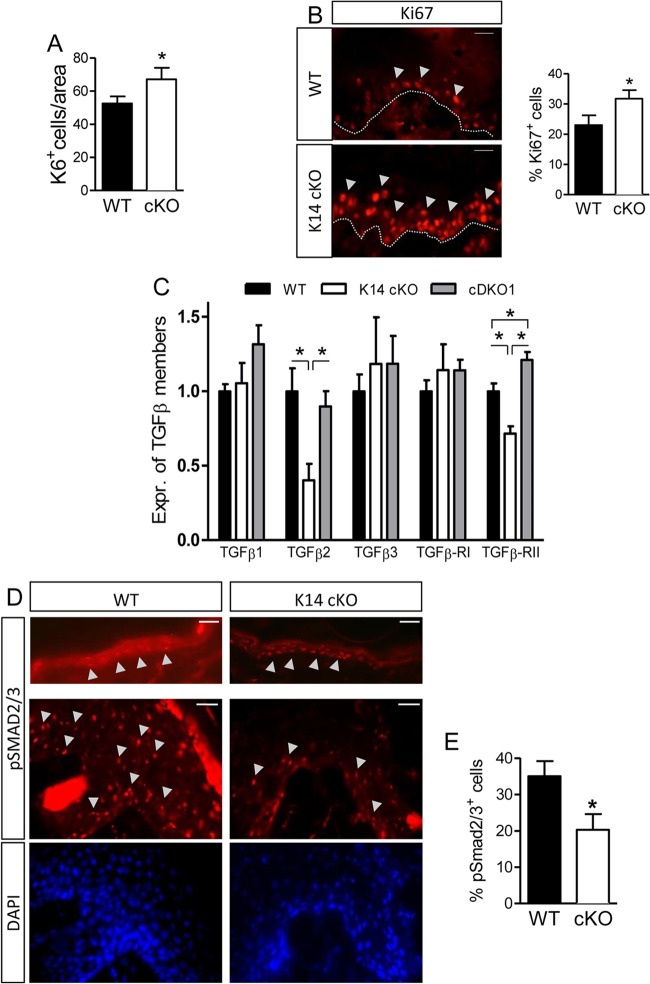
Fig 7.
Enhanced proliferation of keratinocytes in K14 cKO mouse wounds related to the TGFβ pathway. (A) The number of newly formed keratinocytes 5 days after wounding was assessed by quantification of K6+ cells (n = 5 to 8). (B) The proliferation marker Ki67 was stained and found to be expressed in the basal layer (stratum basale) of the keratinocyte tongue of both genotypes. In K14 cKO mice, Ki67+ cells extensively exceeded this layer, suggesting more proliferating cells (n = 7). (C) Isolated keratinocytes were tested for the levels of expression (Expr.) of several TGFβ family members via qPCR (n = 3 to 5). (D) IHC for pSMAD2/3 was performed on unwounded skin sections (top) and 5-day-old wounds (middle). (E) Quantification of pSMAD2/3-positive cells in wounds of both genotypes (n = 5 to 6). Results are given as means ± SEMs. *, P < 0.05. Bars, 25 μm (D, top) and 50 μm (D, middle and bottom).

