Fig 5.
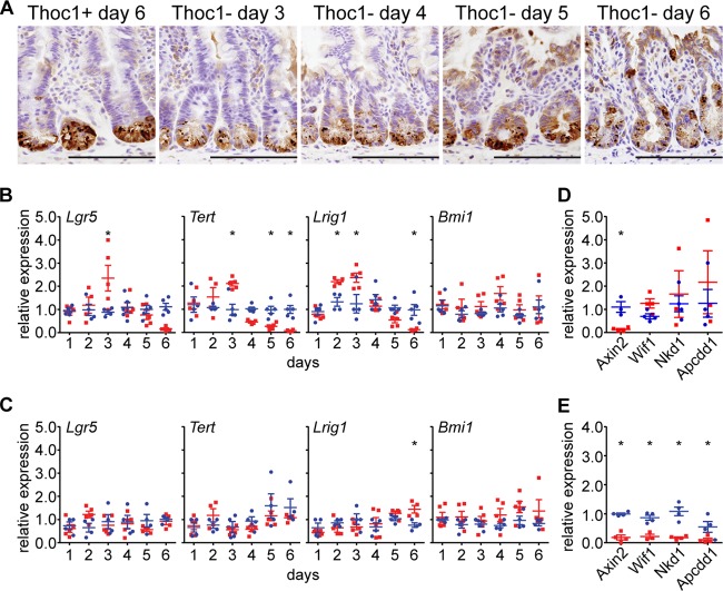
Thoc1 loss disrupts homeostasis of the stem cell niche in the small intestine. (A) Small intestine tissue sections from Thoc1F/F::Rosa26CreERT2 (Thoc1−) or Thoc1+/+::Rosa26CreERT2 (Thoc1+) mice were characterized at the indicated times from the start of tamoxifen treatment by immunostaining for the Paneth cell marker lysozyme. Representative images are shown. Scale bars represent 200 μm. (B) The expression of the indicated stem cell markers in small intestine crypt epithelium from Thoc1F/F::Rosa26CreERT2 (red) or Thoc1+/+::Rosa26CreERT2 (blue) mice was quantitated at the indicated times from the start of tamoxifen treatment by real-time RT-PCR. Each data point is from an individual mouse normalized to a single control mouse at each time point. The bars represent the means and standard errors. Asterisks indicate statistically significant differences between genotypes (t test, P ≤ 0.01). (C) Large intestine tissue was examined for the expression of stem cell marker genes as described for panel B. (D) The expression of the indicated Wnt signaling regulatory genes in small intestine crypt epithelium tissue was assayed at 2 days from the start of tamoxifen treatment as described for panel B. (E) The expression of Wnt regulatory genes was assayed in small intestine crypt epithelium at 5 days from the start of tamoxifen treatment as described for panel B.

