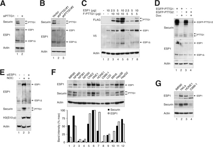
Fig 4.
Reciprocal regulation of the expression of PTTG1 and ESP1. (A) Depletion of PTTG1 reduces endogenous ESP1. HeLa cells were transfected with control or siRNA against PTTG1 (siPTTG1). The cells were harvested 24 h after transfection. Lysates were prepared, and the indicated proteins were detected by immunoblotting. Uniform loading was confirmed by immunoblotting for actin. (B) Depletion of PTTG1 decreases the expression of ESP1. HeLa cells were transfected with control vector or two PTTG1 shRNA-expressing plasmids. A plasmid expressing histone H2B-GFP and a blasticidin resistance gene was cotransfected. After enriching the transfected cells by selection with blasticidin, cell extracts were prepared and subjected to immunoblotting for PTTG1 and ESP1. (C) Ectopic expression of ESP1 increases the expression of PTTG1. HeLa cells were transfected with different amounts of plasmids expressing FLAG-PTTG1 (10 and 5 μg), together with different amounts of ESP1-expressing plasmids (10, 5, and 2.5 μg). Lysates were prepared, and the indicated proteins were detected by immunoblotting. Lysates from vector control (lane 1) and ESP1 alone (lane 2) were loaded as controls. (D) ESP1 is upregulated in PTTG1-overexpressing cells. Cell lines expressing EGFP-PTTG1 or EGFP-PTTG2 were cultured in medium without or supplemented with doxycycline to turn the recombinant protein on or off, respectively. Lysates were prepared, and the expression of PTTG1, PTTG2, and ESP1 was detected by immunoblotting for securin and ESP1. Uniform loading of lysates was confirmed by immunoblotting for actin. (E) Downregulation of ESP1 reduces PTTG1. HeLa cells were transfected with control or siRNA against ESP1. NOC was added for 16 h to trap the cells in mitosis, when the expression of PTTG1 is usually highest. Mitotic cells were collected by mechanical shake off. Lysates were prepared and analyzed by immunoblotting. The increase in histone H3S10 phosphorylation [H3(S10-p)] confirmed the mitotic block. Equal loading of lysates was confirmed by immunoblotting for actin. Control extracts from growing cells were loaded in lane 1. (F) Reciprocal expression of ESP1 and PTTG1 in normal and cancer cell lines. Lysates of various cell lines were prepared and analyzed by immunoblotting for ESP1 and PTTG1. Normal fibroblasts (IMR90) and immortalized nasopharyngeal epithelial (NP460, NP361, and NP550), nasopharyngeal carcinoma (CNE2, HNE1, HONE1, and C666-1), immortalized liver epithelial (LO2 and MIHA), and liver cancer (Hep3B and HepG2) cells were included in the analysis. The ESP1 and PTTG1 signals on the immunoblots were quantified using ImageJ software (National Institutes of Health). (G) Reciprocal expression of ESP1 and PTTG1 in different cell lines during mitosis. The indicated cell lines were treated with nocodazole for 16 h before mitotic cells were isolated by mechanical shake off. Lysates were prepared and analyzed by immunoblotting for ESP1 and PTTG1. Normal fibroblasts (IMR90) and cervical carcinoma (HeLa) and nasopharyngeal carcinoma (HONE1 and C666-1) cells were included in the analysis.

