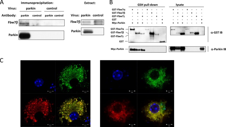
Fig 1.
Parkin associates with Fbw7β in primary neurons. (A) Coprecipitation of exogenous parkin with endogenous Fbw7β in primary neurons. Parkin−/− primary embryonic mouse brain neurons were transduced with either a control adenovirus or an adenovirus expressing N-terminally myc-tagged parkin. Lysates were then immunoprecipitated with a mouse monoclonal antibody specific for parkin or equivalent amounts of mouse IgG. After SDS-PAGE and Western blotting, membranes were probed with Fbw7- and parkin-specific antibodies, respectively. (B) Parkin can form complexes with all Fbw7 isoforms. Retrovirus-transduced HEK293A cell lines stably expressing each of the three Fbw7 isoforms as GST fusions were transfected with a plasmid expressing myc-parkin. Fbw7 complexes were precipitated using glutathione (GSH)-Sepharose beads and analyzed by SDS-PAGE and Western blotting using anti-GST or anti-myc tag antibody. (C) Colocalization of parkin and Fbw7β in primary neurons. GFP-parkin and Fbw7β-DsRed were expressed in primary embryonic mouse brain neurons by lentiviral transduction. Proteins were then detected by laser scanning confocal microscopy. Each of the two panels shows the following: upper left, DNA (DAPI); upper right, GFP-parkin; lower left, Fbw7β-DsRed; lower right, merged image. Scale bar, 5 μm. α, anti; IB, immunoblotting.

