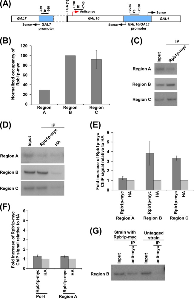
Fig 1.
Analysis of RNA polymerase II association with the antisense start site at the 3′ end of the GAL10 coding sequence. (A) A schematic diagram showing the locations of the primer pairs (regions A, B, and C) at the GAL1, GAL10, and GAL7 loci for the ChIP analysis. Regions A, B, and C are located at the GAL7 core promoter, the 3′ end of the GAL10 coding sequence, and the upstream activating sequence (UAS) at the GAL10-GAL1 bidirectional promoter, respectively. The numbers are presented with respect to the position of the translational stop codon of GAL10. (B) RNA polymerase II is associated with the 3′ end of the GAL10 coding sequence (region B) in dextrose-containing growth medium. A yeast strain expressing the Myc epitope-tagged largest subunit (Rpb1p) of RNA polymerase II was grown in YPD medium to an OD600 of 1.0 at 30°C prior to formaldehyde-based in vivo cross-linking. The ChIP assay was performed as described in Materials and Methods. Immunoprecipitation was carried out using an anti-Myc antibody (9E10; Santa Cruz Biotechnology, Inc.) against Myc-tagged Rpb1p. Immunoprecipitated DNA was analyzed by PCR using the primer pairs encompassing regions A, B, and C as described for panel A. The ratio of the immunoprecipitate over the input in the autoradiogram (termed a ChIP signal) was measured. The maximum ChIP signal was set to 100, and other ChIP signals were normalized with respect to 100 (represented as normalized or relative occupancy). Normalized occupancy was plotted in the form of a histogram. (C) Autoradiograms for the ChIP data presented in panel B. IP, immunoprecipitation. (D) Analysis of RNA polymerase II at the 3′ end of the GAL10 coding sequence (region B), GAL7 core promoter (region A), and GAL1-GAL10 UAS (region C) along with anti-HA as a nonspecific antibody in dextrose-containing growth medium. A yeast strain expressing Myc-tagged Rpb1p was grown in YPG medium to an OD600 of 0.8 at 30°C and then transferred to YPD medium for 3 h prior to cross-linking. (E) The ratio of the ChIP signal of Myc-tagged Rpb1p to anti-HA (i.e., fold increase of Rpb1p-Myc ChIP signal relative to HA) in panel D is plotted in the form of a histogram. A ratio of 1 indicates no association of RNA polymerase II. (F) Analysis of association of RNA polymerase II with the RNA polymerase I gene (Pol I) relative to anti-HA in dextrose-containing growth medium. The ratio of the ChIP signal of Myc-tagged Rpb1p to anti-HA was plotted in the form of a histogram. (G) The ChIP analysis of RNA polymerase II association at the 3′ end of the GAL10 coding sequence (region B) in the yeast strain expressing Rpb1p with or without a Myc epitope tag. Yeast strains were grown as described for panel D. Immunoprecipitation was carried out using an anti-Myc antibody.

