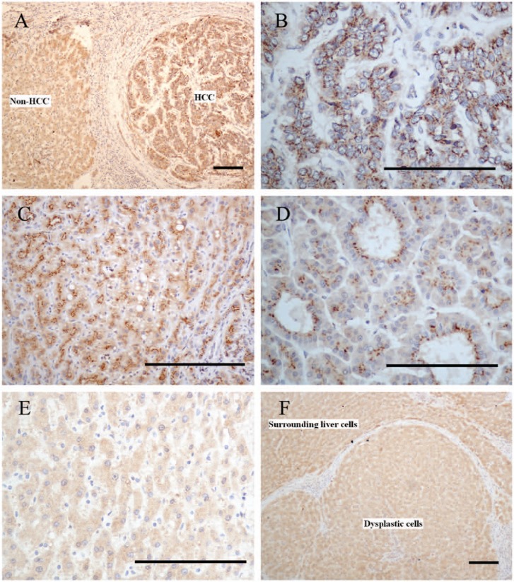
Figure 2.
Different GP73 immuno-staining patterns in hepatocellular carcinoma (HCC) cells and non-HCC cells. (A) The immunostaining pattern of GP73 in HCC cells showed a diffuse coarse-block pattern. In contrast, the surrounding non-HCC cells showed a diffuse fine-granular pattern. (B) The coarse blocks were perinuclear. (C) The coarse blocks were concentrated in a certain part of the cytoplasm that gave a cluster- or cord-like appearance. (D) The coarse blocks were concentrated near the lumen of glandular structures. (E) The surrounding non-HCC cells showed a diffuse fine-granular pattern in the cytoplasm. (F) A case of a high-grade dysplastic nodule, which showed a diffuse fine-granular pattern with no difference in intensity between dysplastic cells and surrounding liver cells. Bars = 100 µm.

