
Figure 3.
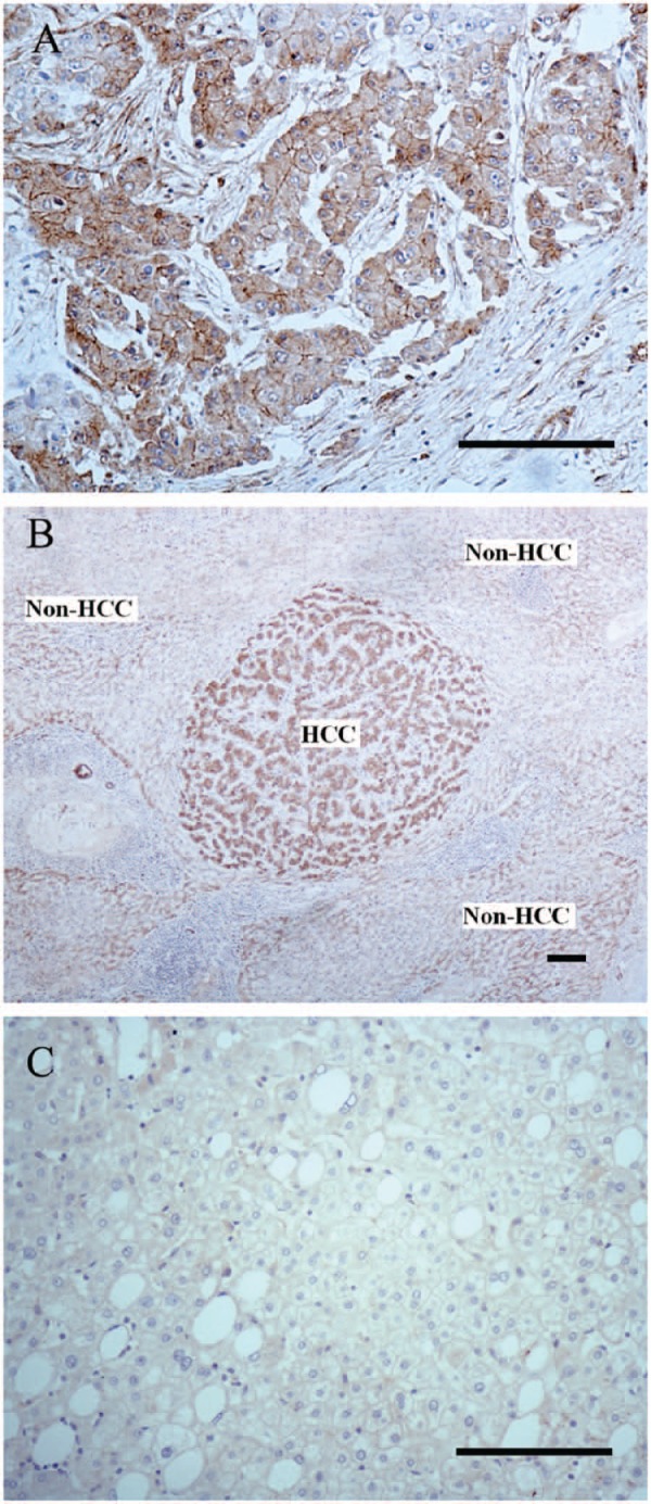
GPC3 immunostaining patterns in hepatocellular carcinoma (HCC) cells and non-HCC cells. (A) The immunoreactive pattern of GPC3 in HCC cells showed a diffuse cytoplasmic and membrane-associated pattern. (B) The intensity of GPC3 immunoreaction in HCC cells was greater than that of surrounding non-tumor cells in a case of well-differentiated HCC. (C) GPC3 staining was negative in a case of focal nodular hyperplasia. Bars = 100 µm.