Figure 2.
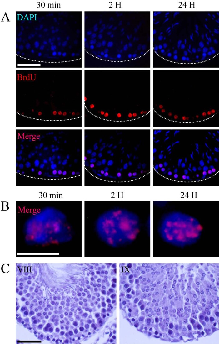
Light microscope micrographs showing type II labeling. (A) Panoramic view of transversal sections of seminiferous tubules showing the distribution of BrdU-labeled cells. The nuclear chromatin is evidenced by DAPI staining. Immunodetection of BrdU shows scattered foci, dispersed throughout the nuclear space. These positive sites were present at all incorporation times. The positive cells are those closest to the basal membrane of the seminiferous tubule and correspond to phases VIII and IX of the spermatogenic cycle (C). (B) High magnification shows the specific distribution pattern of BrdU in the nuclei. The localization of the positive staining is associated with loose and compact chromatin. Dotted lines show the border of the seminiferous tubule. Scale bars: (A) 50 microns; (B) 5 microns; (C) 30 microns.

