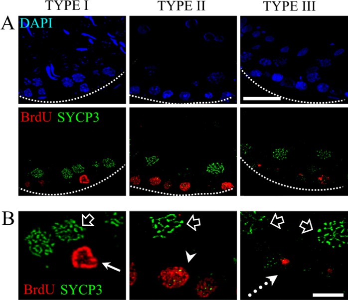
Figure 6.
Double immunolocalization of BrdU and the SYCP3 protein. (A) Image displaying the three different patterns of BrdU incorporation. Type I corresponds to cells in the mitotic S phase that are not positive for the meiotic protein SYCP3, whereas the cells with pattern types II and III are positive for the SYCP3 protein. (B) High magnification of the three patterns described. The arrow shows a cell with pattern type I, which is negative for the SYCP3 protein. The arrowhead points to a double-labeled cell that is positive for SYCP3 and BrdU incorporation and corresponds to pattern type II. The dotted arrow shows a cell with pattern type III, which is simultaneously positive for the SYCP3 protein. Empty arrows point to cells that are positive for the SYCP3 protein and form filaments; these cells are in the pachytene stage. Dotted lines show the border of the seminiferous tubule. Scale bars: (A) 30 microns; (B) 10 microns.

