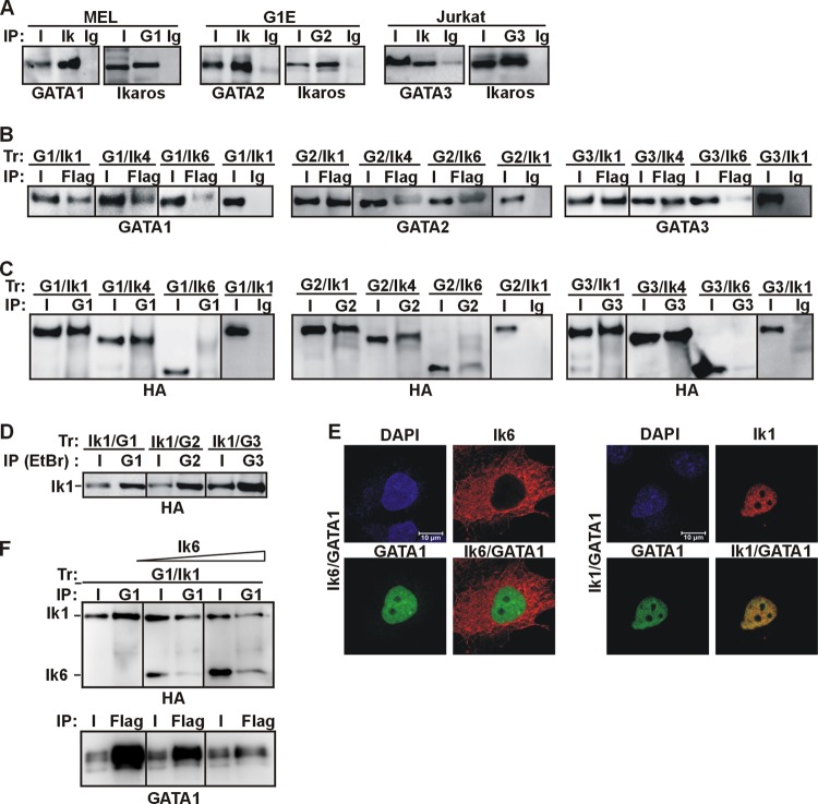
Fig 1.
Ikaros-GATA protein interactions in vivo. (A) The Ik-GATA interaction in hematopoietic cell lines. Total cell lysates of MEL, G1E, or Jurkat cells were used for protein co-IP with Ik, GATA1 (G1), GATA2 (G2), or GATA3 (G3) antibodies or control Ig (Ig). Input samples (I) represented 2% of total lysates. The co-IP samples were analyzed by Western blotting with GATA or Ik antibodies. (B and C) Total cell lysates of COS-7 cells transfected with expression vectors encoding various combinations of GATA1 (G1), GATA2 (G2), GATA3 (G3), Ik1-FH (Ik1), Ik4-FH (Ik4), or Ik6-FH (Ik6) were immunoprecipitated with Flag or GATA antibodies or control Ig (mouse Ig for Flag co-IP; rat Ig for GATA1 co-IP; mouse Ig for GATA2 and GATA3 co-IP). The co-IP samples were analyzed by Western blotting with GATA or HA antibodies. (D) Total cell lysates of COS-7 cells transfected with expression vectors encoding Ik1-FH and GATA1 (Ik1/G1), Ik1-FH and GATA2 (Ik1/G2), or Ik1-FH and GATA3 (Ik1/G3) were immunoprecipitated with GATA1, GATA2, or GATA3 antibodies in lysis buffers containing 1 mM DTT, 2 mM β-mercaptoethanol, and 50 μg/ml ethidium bromide. Co-IP samples were analyzed by Western blotting with HA antibody. (E) Confocal IF of COS-7 cells expressing Ik1-FH or Ik6-FH together with GATA1. GATA1 (green signal) was detected with rat anti-GATA1 and FITC-conjugated anti-rat antibodies; Ik (red signal) was detected with mouse anti-HA and Texas Red (TR)-conjugated anti-mouse antibodies. Representative COS-7 cells are shown where Ik1-GATA1 colocalized, as yellow signals in merged images. (F) In vivo squelching assay. Total cell lysates of COS-7 cells transfected with Ik1-FH and GATA1 and an increasing concentration of Ik6-FH were immunoprecipitated either with GATA1 or Flag antibodies. Co-IP samples were analyzed by Western blotting with HA or GATA1 antibodies.

