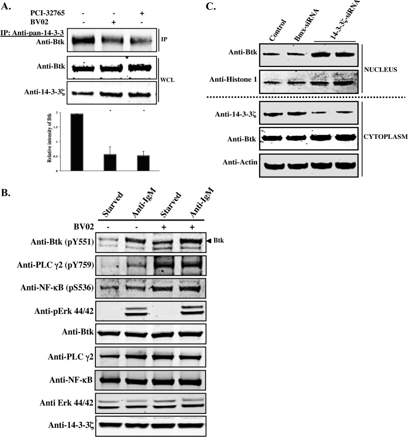
Fig 4.
Inhibition of Btk and 14-3-3ζ. (A) Primary mouse B cells were treated with the Btk inhibitor PCI-32765 and the nonpeptide 14-3-3 inhibitor BV02 and incubated for 24 h. Control samples were incubated with DMSO. WCLs were processed for Western blot analysis. Quantitative analysis of Btk bands was performed by densitometry analysis using Image J software. The bars represent the means and SE of two independent experiments. *, P < 0.015. (B) Namalwa cells were starved or activated using anti-IgM (20 μg/ml) for 15 min in the presence or absence of BV02. WCLs were processed for Western blotting using phospho-specific antibodies against Btk (pY551), PLCγ2 (pY759), NF-κB (pS536), or pErk p44/42 (Thr202/Tyr204). (C) siRNA-mediated depletion of endogenous 14-3-3ζ in Namalwa cells. Cells were transfected with a 14-3-3ζ siRNA (two independent experiments) or with control siRNA targeting Bmx, another Tec family member. The siRNA-treated cells were collected 48 h after transfection. Western blot analysis was performed using lysates prepared from nuclear and cytoplasmic fractions. The purity of the nuclear and cytoplasmic fractions was verified by probing with antibodies against H1 histone and β-actin, respectively.

