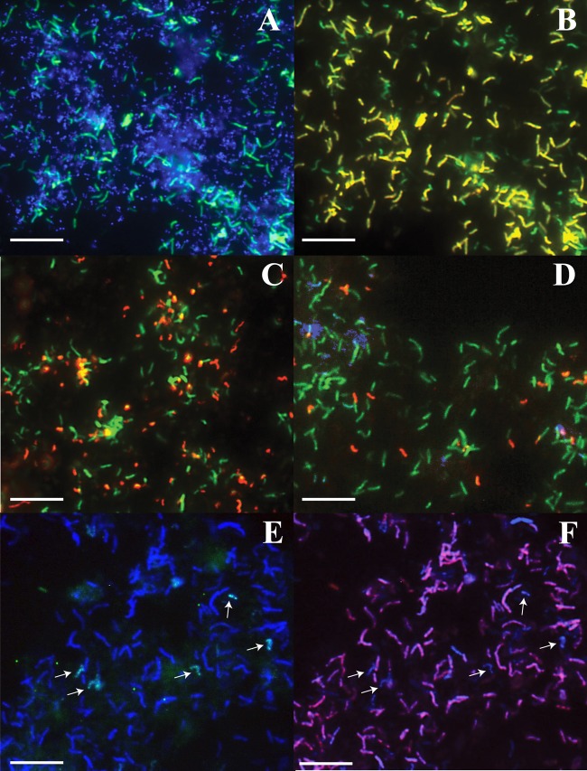
Fig 1.
Fluorescence in situ hybridization images of UBA BS biofilms. (A) Sample Aug07, showing DNA (blue; most small dots are Archaea) and general bacteria (green). (B) Sample Aug07, showing general bacteria (green) and Leptospirillum group IV UBA BS (yellow). (C) Sample Jun08, showing general bacteria (green) and L. ferrodiazotrophum (red). (D) Sample Aug07, showing general bacteria (green), L. ferrodiazotrophum (red), and Leptospirillum group II 5wayCG type (blue). (E) Sample Island 2, showing general bacteria (blue), L. ferrodiazotrophum (green/light blue). (F) Sample Island 2, showing general bacteria (blue) and Leptospirillum group IV UBA BS (purple/pink). The images in panels A and B were taken from the same field of view, as were the images in panels E and F. The white arrows indicate Leptospirillum group III cells. Bars, 1 μm.

