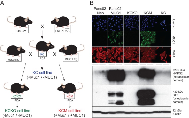
Fig 1.
Mouse PDA cell lines used in this study. (A) KC mice producing KRASG12D-driven spontaneous PDAs (KC cells) were crossed with mice expressing human MUC1 (MUC1.Tg) or MUC1 null (MUC1KO) to generate the MUC1-positive KCM or MUC1-null KCKO cell lines, respectively. (B) MUC1 expression profile of PDA cell lines. For immunofluorescence (IF) analysis using confocal microscopy, cells were analyzed using HMFG2 antibody to detect the extracellular domain of human MUC1 and FITC-conjugated secondary antibody. Hoechst dye was used to stain for the nucleus, and wheat germ agglutinin (WGA) was used to stain the plasma membrane. For Western blot analysis, total cell lysates were separated by SDS-PAGE and then analyzed by Western blotting with HMFG2 antibody or CT2 antibody to detect the transmembrane domain of human MUC1. Western blotting using β-actin antibody was used as a loading control.

