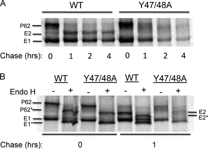
Fig 3.

Envelope protein biosynthesis in WT- and Y47/48A-infected cells. BHK cells were electroporated with either WT or mutant RNA and incubated at 37°C for 6 h, pulse-labeled for 30 min with [35S]methionine-cysteine, and then chased for the indicated times. The cell lysates were immunoprecipitated with a polyclonal antibody to the SFV envelope glycoproteins. Samples were analyzed directly by SDS-PAGE (A) or following digestion with Endo H (B). p62*, E1*, and E2* illustrate the new positions of the proteins after Endo H digestion. Representative examples of two independent experiments are shown.
