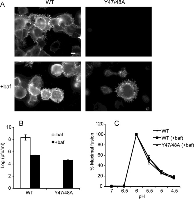
Fig 6.

Assembly properties of Y47/48A after neutralization of exocytic pH. BHK cells were electroporated with WT or mutant viral RNA and incubated for 2 h at 37°C. Medium with (+) or without (−) bafilomycin (baf) was added, and the incubation was continued for 8 to 12 h. (A) After 12 h, the surface expression of E1 was visualized as described in the legend of Fig. 4. Fluorescence microscopy images were acquired with the same exposure times. Scale bar, 10 μm. (B) After 8 h, medium samples were collected, and titers were determined by plaque assay on BHK cells. (C) Virus stocks from the experiment described in panel B were also used to determine the pH dependence of virus fusion with the plasma membrane of BHK cells, as described in Materials and Methods. The images in panel A are representative of three independent experiments. Data in panels B and C represent the average and standard deviation of three independent experiments.
