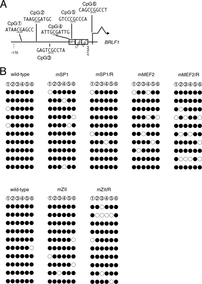
Fig 9.
CpG DNA methylation of the BRLF1 promoter in mutant LCLs. (A) Schematic illustration of the BRLF1 promoter. The distribution of six CpG dinucleotides analyzed in panel B is shown with circled numbers. cis-element motifs and BRLF1 coding region are also depicted. (B) CpG DNA methylation of the BRLF1 promoter in B cells. DNA from LCLs latently infected with wild-type or mutant recombinant EBV-BAC was subjected to bisulfite modification, followed by sequencing. Filled circles, methylated; open circles, unmethylated.

