
Fig 6.
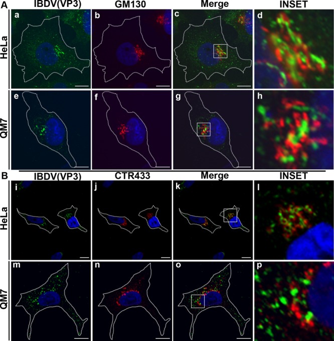
Juxtanuclear localization of IBDV VP3. Infected HeLa and QM7 cells were processed for indirect immunofluorescence analysis at 36 h p.i. with anti-VP3 (1:500) and the corresponding Alexa 488-conjugated secondary antibodies. Double labeling was performed with anti-GM130 (A) or anti-CTR433 (B) and the corresponding Cy3-conjugated secondary antibodies. The insets show the strong association of VP3 (green signal) and the Golgi markers (red signal) at the perinuclear region. The nuclei were stained with Hoechst stain (blue). Images are representative of three independent experiments. Scale bars represent 10 μm.