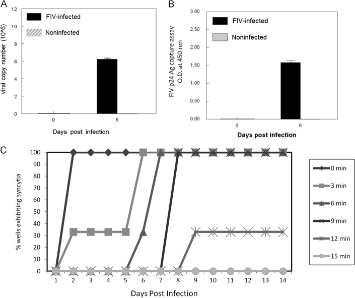
Fig 1.
Validation of CD4+ CD25+ in vitro infection methods. Purified feline CD4+ CD25+ cells were infected with the NCSU1 isolate of FIV at an MOI of 2.5. Cells and culture supernatant were harvested at 6 days postinfection and analyzed for virus copy by real-time RT-PCR (A) and p24 antigen (Ag) by ELISA (B). Each bar represents the mean ± standard error of the mean (SEM) of 3 replicates. O.D., optical density. (C) UV inactivation of FIV was confirmed using the FIV-susceptible feline T cell line FCD4-E. Virus was exposed to 1 μJ of UV light for various times and then added to FCD4-E cells at an MOI of 2. Five cultures per UV inactivation time were observed daily up to 14 days for the formation of syncytia.

