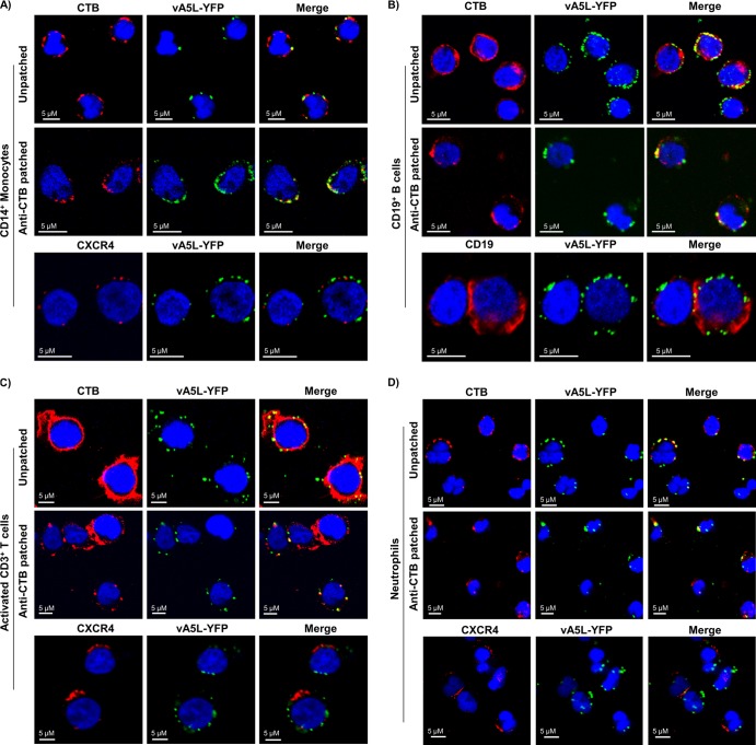
Fig 2.
VV binding to lipid rafts on the surface of PHL subsets. Isolated monocytes (A), B cells (B), activated T cells on day 3 of activation (C), and neutrophils (D) were patched or unpatched and then subjected to staining with CTB conjugated with Alexa Fluor 647 (red), anti-human CXCR4 Ab conjugated with Alexa Fluor 647 (red) for monocytes, activated T cells, and neutrophils, or anti-human CD19 Ab conjugated with Alexa Fluor 647 (red) for B cells. All cell types were then incubated with vA5L-YFP (green) at an MOI of 10 under binding conditions, fixed with 2% PFA, and adhered to poly-l-lysine-coated coverslips. Coverslips were mounted with mounting medium containing DAPI (blue) and analyzed by confocal microscopy. Scale bars represent 10 μM. The data represent the results of VV binding to lipid rafts on PHL subsets from 6 blood donors.

