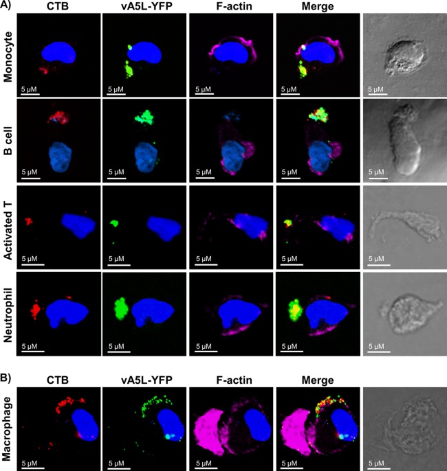
Fig 3.
VV binding to lipid rafts enriched in uropods of polarized PHL subsets. (A) VV binding to lipid rafts enriched in uropods of polarized monocytes, B cells, activated T cells on day 3 of activation, and neutrophils. These cell types were treated with GM-CSF, SDF-1, anti-CD44-coated plates, and bacterial peptide fNLPNTL, respectively, to induce cell polarization. Polarized cells were subsequently fixed with 2% PFA and stained with CTB conjugated with Alexa Fluor 647 (red), phalloidin conjugated with Alexa Fluor 546 to stain actin filaments (pink), and DAPI (blue). Cells were then subjected to VV binding with vA5L-YFP (green) and confocal microscopy analysis. (B) VV binding to lipid rafts enriched in uropods of polarized macrophages that were differentiated from monocytes after 7 days of culture in vitro in the presence of GM-CSF. Scale bars represent 10 μM. The data represent the results of VV binding to these polarized cell types derived from 6 blood donors.

