Fig 4.
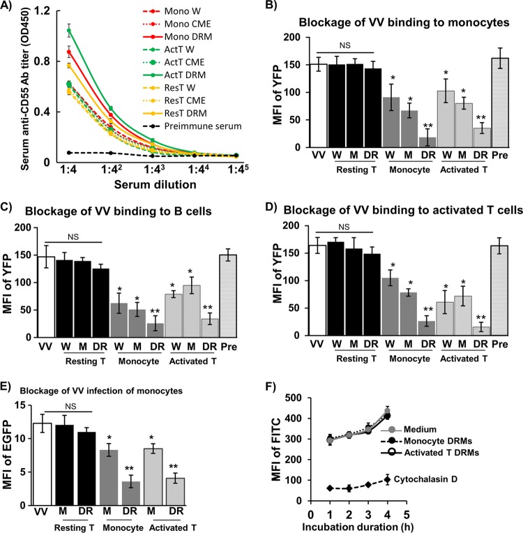
Blockage of VV binding and infection of PHL subsets by immunosera. (A) Titers of anti-human CD55 Abs in immunosera raised against DRMs, CMEs, or whole cells from monocytes, resting T cells, or activated T cells were determined using an ELISA. Immunosera diluted at 1:10 with PBS were used to block VV binding to monocytes (B), B cells (C), and activated T cells (D). (E) Immunosera diluted at 1:10 with PBS were used to block VV infection of monocytes. (F) Effects of immunosera diluted at 1:10 with PBS and cytochalasin D on monocyte endocytosis of FITC-latex beads. The MFI of YFP (FITC for latex beads) or EGFP represented VV binding or infection intensity to PHL subsets of 6 blood donors. Data were compared using Tukey's ANOVA assay. Mono W, whole monocytes; Mono CME, monocyte crude membrane extracts (CMEs); Mono DRM, monocyte detergent-resistant membranes (DRMs); ActT W, whole activated T cells on day 3 of activation; ActT CME, activated T cell CMEs; ActT DRM, activated T cell DRMs; ResT W, whole resting T cells; ResT CME, resting T cell CMEs; ResT DRM, resting T cell DRMs; Pre, preimmunization sera. Statistical analysis was used to compare each experimental condition with medium (control). NS, not significant; *, P < 0.05; **, P < 0.01.

