
Fig 4.
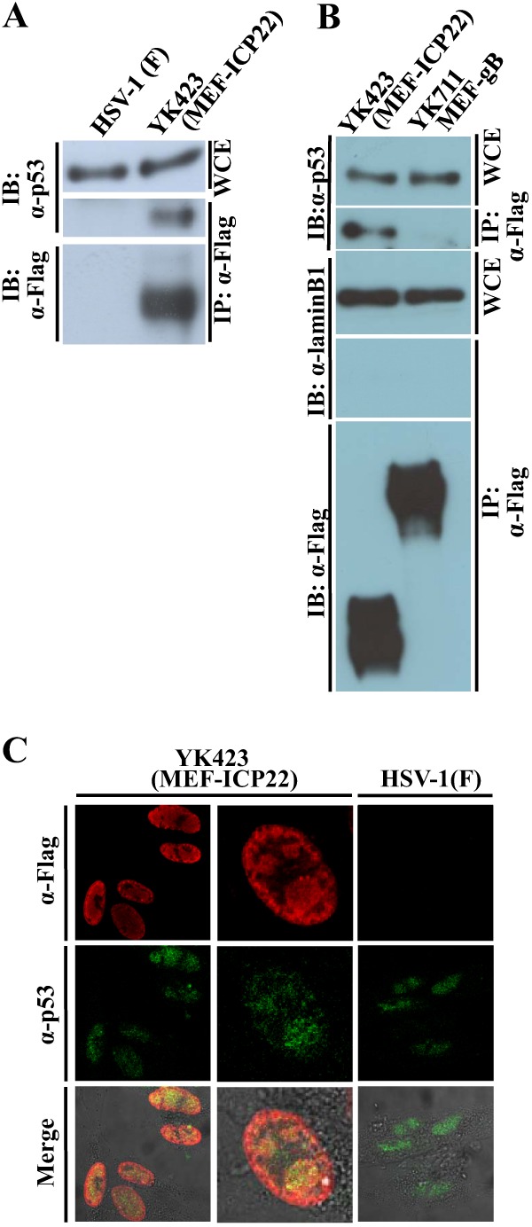
Interaction of ICP22 with p53. (A) HEL cells infected with wild-type HSV-1F (lane 1) or YK423 (MEF-ICP22) (lane 2) at an MOI of 5 for 9 h were harvested, immunoprecipitated (IP) with anti-Flag antibody (α-Flag), and analyzed by immunoblotting (IB) with anti-p53 antibody (α-p53) or anti-Flag antibody. WCE, whole-cell extract. (B) HEL cells infected with wild-type HSV-1F (lane 1) or YK711 (MEF-gB) (lane 2) at an MOI of 5 for 9 h were harvested, immunoprecipitated (IP) with anti-Flag antibody (α-Flag), and analyzed by immunoblotting (IB) with anti-p53 antibody (α-p53), anti-laminB1 (α-laminB1), or anti-Flag antibody. WCE, whole-cell extract. (C) HEL cells infected with YK423 (MEF-ICP22) or wild-type HSV-1F at an MOI of 10 for 9 h were fixed, permeabilized, stained with anti-Flag and anti-p53 antibodies, and examined by confocal microscopy. Upper and middle columns and the lower column show protein fluorescence and simultaneous acquisition of protein fluorescence and digital interference contrast, respectively.