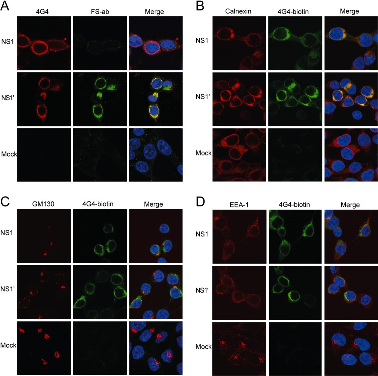
Fig 2.
Cellular localization of plasmid-expressed NS1′ and NS1. (A) Immunofluorescence analysis showing production of NS1 and NS1′ using 4G4 (which stains both NS1 and NS1′) and FS-Ab (an NS1′-specific antibody) in transfected HEK293T cells. (B to D) Immunofluorescence analysis showing colocalization of plasmid-expressed NS1 and NS1′ with the ER (B), the Golgi apparatus (C), and endosomes (D). Transfected HEK293T cells were stained with antibodies to appropriate cell markers (calnexin, GM130, and EEA-1, respectively) and biotinylated anti-NS1 (4G4).

