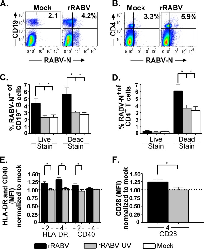
Fig 7.
Human B and T cells from healthy nonimmunized donor PBMCs are infected by rRABV in vitro and upregulate the markers of antigen presentation and costimulation, HLA-DR, CD40, and CD28. Freshly isolated PBMCs were cultured at a density of 106 cells/ml and mock infected or infected with either rRABV or rRABV-UV at an MOI of 5 for 4 days. Cells were harvested and stained for B and T cell lineage marker expression, as well as costimulatory activation markers, for analysis by flow cytometry. (A) Representative gating strategy for live (LIVE/DEAD stain low) human cells stained for CD19 and RABV-N from mock-infected (left) or rRABV-infected (right) cultures. (B) Representative gating strategy for dead (LIVE/DEAD stain high) human cells stained with CD4 and RABV-N from mock-infected (left) or rRABV-infected (right) cultures. (C) Percentages of live and dead CD19+ B cells staining RABV-N+ 4 days postinfection in vitro from cultures infected with rRABV or rRABV-UV or mock infected. (D) Percentages of live and dead CD4+ T cells staining RABV-N+ 4 days postinfection in vitro from cultures infected with rRABV or rRABV-UV or mock infected. (E) MFI of HLA-DR and CD40 staining on days 2 and 4 postinfection of RABV-N+ CD19+ B cells from PBMC cultures infected with rRABV (black bars) or rRABV-UV (gray bars), normalized to the MFI of HLA-DR or CD40 on CD19+ B cells from mock-infected cultures. (F) MFI of CD28 staining on RABV-N+CD4+ T cells from PBMC cultures infected with rRABV (black bar) or rRABV-UV (gray bar), normalized to the MFI of CD28 on CD4+ T cells from mock-infected cultures. To compare two groups of data, we used an unpaired, two-tailed Student's t test (*, P < 0.05).

