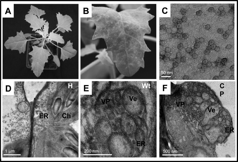
Fig 4.
Symptom phenotypes and membrane rearrangements induced by wt BMV and its CP in C. quinoa leaves. (A) Healthy leaves; (B) a leaf systemically infected with BMV; (C) TEM image of negatively stained BMV virions purified from systemically infected C. quinoa. TEM images showing unmodified membranes in a healthy leaf (D) or membrane modifications induced by wt BMV (E) or ectopically expressed wt CP (F). Ch, chloroplast; ER, endoplasmic reticulum; VP, vesicle pocket; Ve, vesicle.

