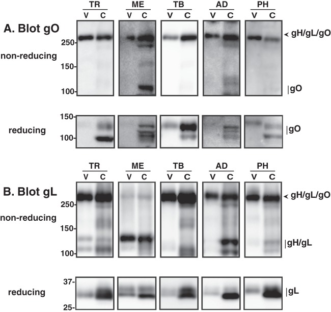
Fig 7.
Comparative analysis of gH/gL/gO complexes from different strains of HCMV. Extracts of HFF cells (C) infected with BAC-derived HCMV TR, Merlin, TB40/e, AD169, or PH or virions (V) collected from the culture supernatant were separated by nonreducing or reducing SDS-PAGE, transferred to PVDF membranes, and probed with anti-gO (A) or anti-gL (B) antibodies. For gO blots, anti-TBgO antibodies were used for TR, TB, and PH samples, anti-MEgO antibodies were used for ME samples, and anti-ADgO antibodies were used for AD samples. Arrowheads mark bands corresponding to the disulfide-linked gH/gL/gO trimer, and vertical lines indicate bands that correspond to gO, gL, or the disulfide-linked gH/gL heterodimer. Mass markers (kDa) are shown on the left.

