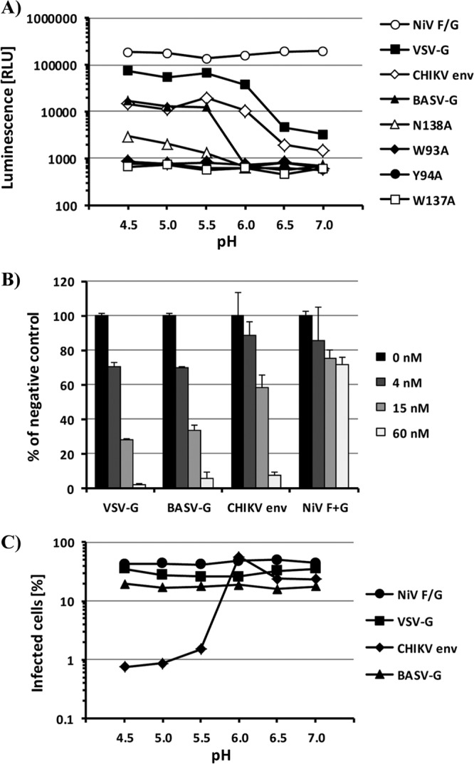
Fig 4.

Activation of BASV-G fusion activity is pH dependent and reversible and occurs in the late endosome. (A) Cell-cell fusion mediated by the viral glycoproteins NiV F/G, CHIKV env, VSV-G, BASV-G, and the BASV-G W93A, Y94A, W137A, and N138A fusion loop mutants. Vero cells expressing the alpha subunit of β-galactosidase and 293T cells expressing the omega subunit of β-galactosidase and one of the glycoproteins were mixed and allowed to fuse at the indicated pH values. β-Galactosidase activities in cell lysates were determined at 5 h postfusion by using the chemiluminescence-based GalactoLight assay system. The experiment was performed in triplicate, and a representative experiment of a total of five experiments is shown. RLU, relative light units. (B) Vero cells were incubated with the indicated concentrations of bafilomycin A prior to infection with VSVΔG-GFP pseudotypes bearing VSV-G, BASV-G, CHIKV env, or NiV F/G. The level of infection was determined as the percentage of GFP-expressing cells at 24 h postinfection and is shown as a percentage of infection in the absence of bafilomycin A. The experiment was performed in duplicate, and a representative experiment of a total of four experiments is shown. Error bars indicate standard deviations. (C) VSVΔG-GFP pseudotypes bearing VSV-G, BASV-G, CHIKV env, or NiV F/G were exposed to the indicated pH values for 30 min before neutralization and infection of Vero cells. The level of infection was determined as a percentage of GFP-expressing cells at 24 h postinfection. The experiment was performed in duplicate, and a representative experiment of a total of three experiments is shown.
