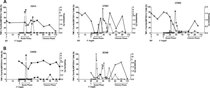
Fig 1.
Correlation of peripheral blood parasitemia and myeloid dendritic cell (mDC) TLR4 responsiveness. TLR4 responsiveness (solid line) and parasitemia (dashed line) are shown for individual animals that were coinfected with SIV and P. fragile (A) or infected only with P. fragile (B). TLR4 responses were measured after in vitro stimulation of peripheral blood mononuclear cells with 1 μg/ml lipopolysaccharide (LPS) and subsequent multicolor flow-cytometric analysis. The percentage of TNF-α+ cells in the total mDC population (Lin− CD11c+ HLA-DR+) is shown. Parasitemia is reported as the percentage of infected red blood cells.

