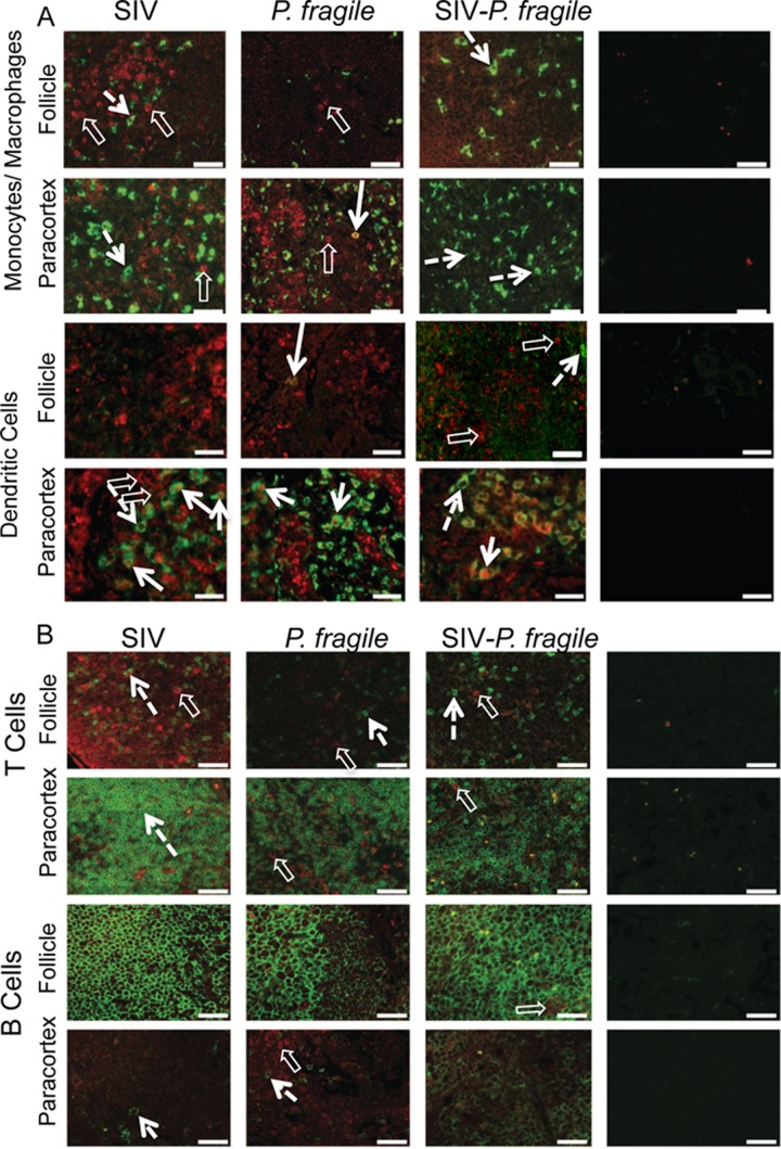
Fig 6.
IL-10 expression in axillary lymph nodes. Immunofluorescence was used to determine the phenotype of IL-10-producing cells in the paracortex (T cell zone) and follicle (B cell area) of axillary lymph nodes. Representative tissue sections of SIV-, P. fragile-, and SIV-P. fragile-infected animals at necropsy are shown. Images show sections stained for IL-10 (red) and Mo/Mph or mDC markers (A) or sections stained for IL-10 and T or B cell markers (B). Single- and double-positive cells are identified as described in the legend to Fig. 4.

